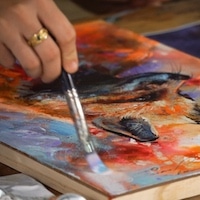
Neurobiologist Igor Siwanowicz marries science and art with these incredibly-detailed images taken with a confocal laser-scanning microscope. Using the hi-tech device, he uncovers the invisible world of small creatures such as barnacles, moths, and beetles. Siwanowicz, who has won awards for his photography, works at the Howard Hughes Medical Institute’s Janelia Farm Research Campus and uses this special microscope as creative escape from his stressful work. “You need creative outlets,” he shares. “Especially photography, where the magic happens in a split second where you press the shutter. You're not dwelling in the past, or thinking about the future. You're in the moment. It was very therapeutic for me.”
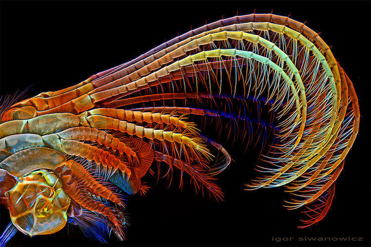
The confocal laser-scanning microscope has the ability to capture an unparalleled amount of detail in comparison to a lens-based microscope. It works by taking multiple photographs at different levels of focus, reconstructing them into a single high-resolution, depth-enhanced image. The surreal colors are owed to fluorescent dyes used to illuminate the samples, making these minuscule creatures seem extraterrestrial.
Siwanowicz's images highlight the beautiful complexity of nature. Even the smallest of creatures contains millions of details, contrasted all the more by the glowing colors of the photographs. The element of surprise is part of the fascination for Siwanowicz, as not even he is sure of what he will discover until the image is finalized. Discover more of his microscope photography here.
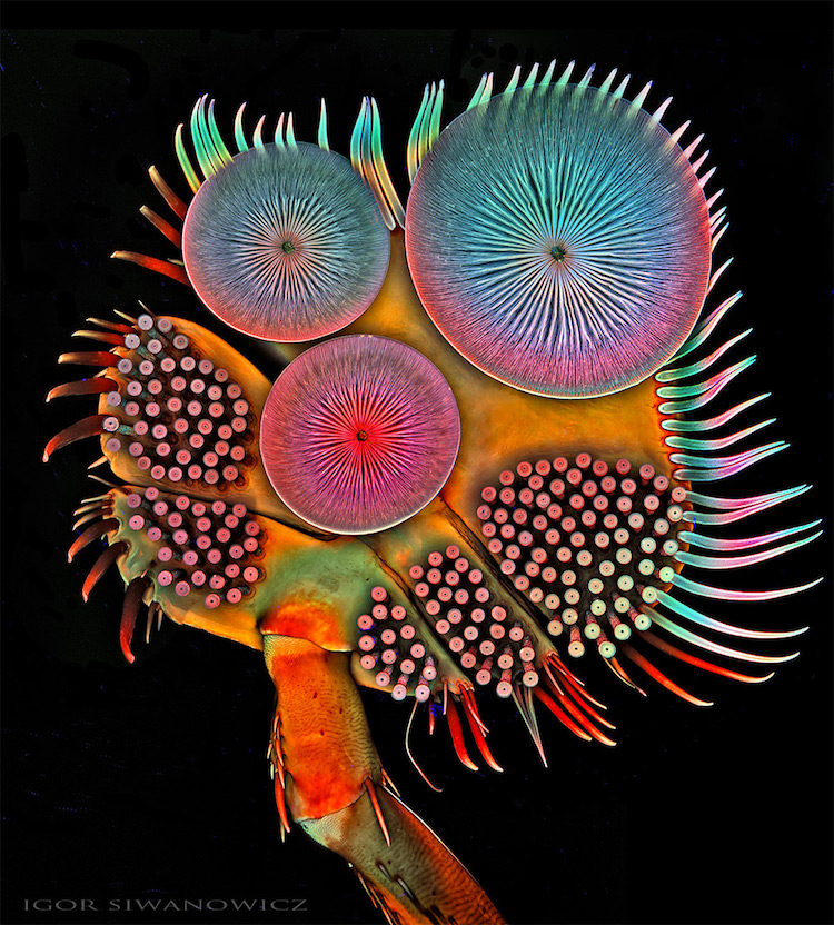
Above image: Acilius diving beetle male front tarsus (foot) 100x
Barnacle
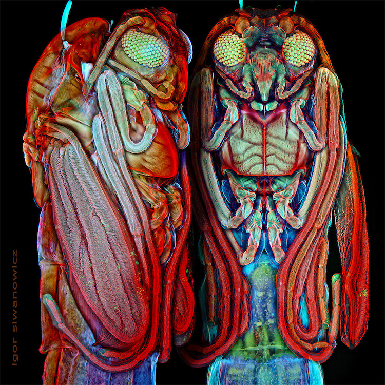
Midge Pupa
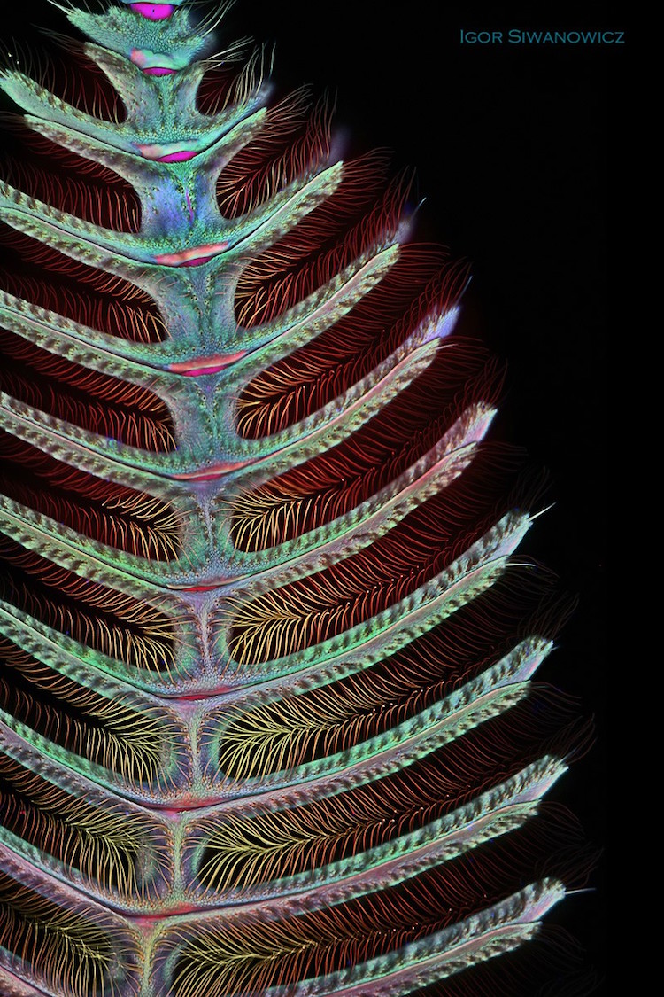
Moth antennae
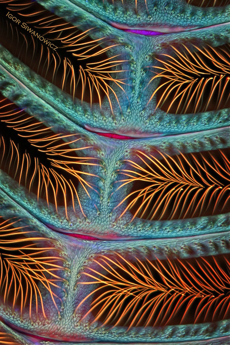
Moth antennae (detail)
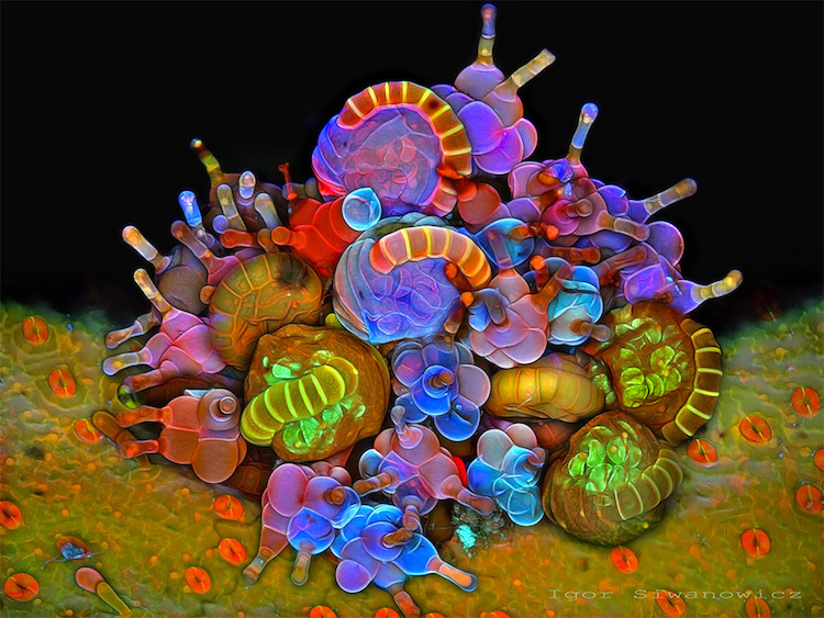
Paraphyses and Sporangia
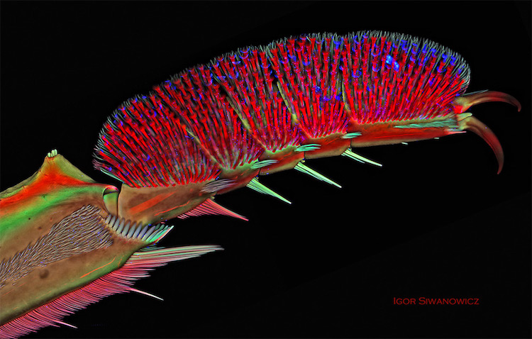
Front leg of whirligig beetle
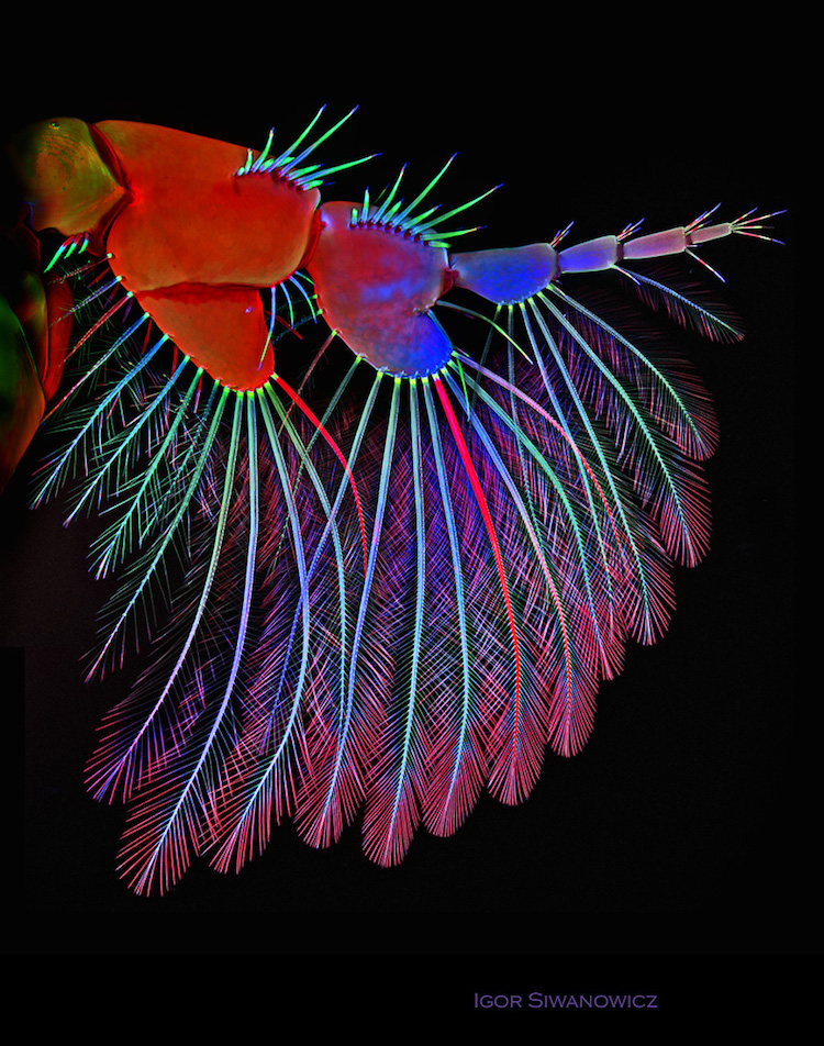
Isopod appendage
Igor Siwanowicz: Website | Facebook
via [Colossal]
All images via Igor Siwanowicz