If you find yourself in Virginia's Dulles International Airport through November of this year, you might see the exhibition Life: Magnified on the walls. It features scientific images showing cells and other scenes of life magnified by as much as 50,000 times. The brilliantly-colored and beautifully-textured works depict parts of the human body such as the brain and blood cells, in addition to showing us what a fruit fly and lone star tick look like up close.
These incredible magnifications don't necessarily look like what they actually are; they could easily be mistaken for an abstract drawing or painting. Their vibrant hues aren't naturally occurring, however, and are the result of chemical dyes or design programs that allow scientists to study selected structures within a cell.
You can view more of these images through the exhibition's website and also learn about how they affect us and our world. Life: Magnified was organized by the National Institute of General Medical Sciences, the American Society for Cell Biology, and the Metropolitan Washington Airports Authority's Arts Program.
Above: Anglerfish Ovary Cross-Section. Image credit: James E. Hayden, The Wistar Institute, Philadelphia, Pa.
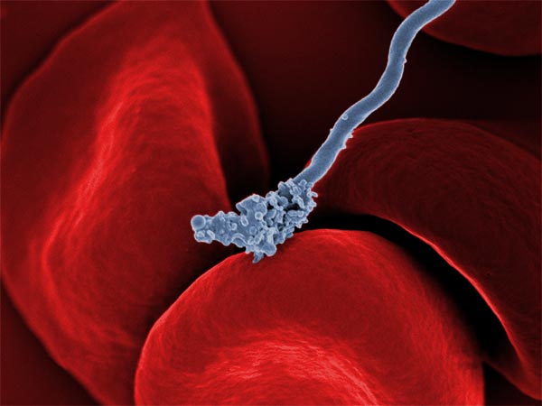 Relapsing Fever Bacterium (Gray) on Red Blood Cells
Image credit: Tom Schwan, Robert Fischer and Anita Mora, National Institute of Allergy and Infectious Diseases, National Institutes of Health
 Human Liver Cell (Hepatocyte)
Image credit: Donna Beer Stolz, University of Pittsburgh
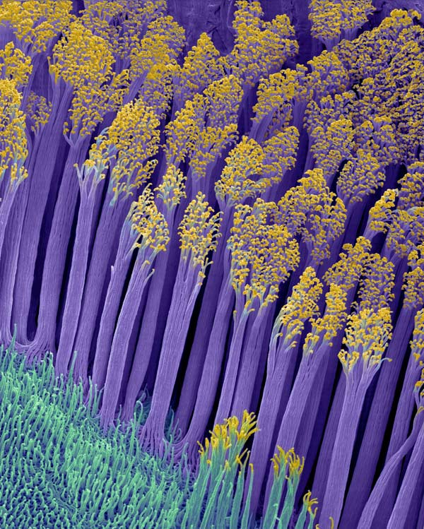 Gecko Lizard Toe Hairs
Image credit: Dennis Kunkel, Dennis Kunkel Microscopy, Inc.
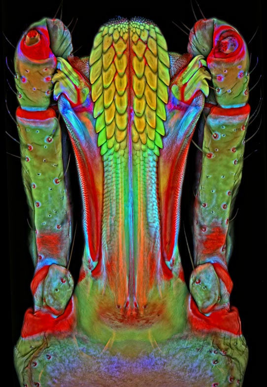 Mouth Parts of a Lone Star Tick
Image credit: Igor Siwanowicz, Janelia Farm Research Campus, Howard Hughes Medical Institute, Ashburn, Va.
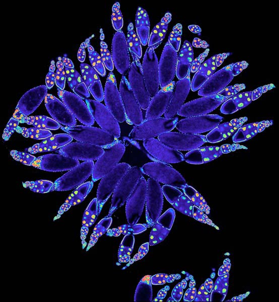 Fruit Fly Ovary
Image credit: Hogan Tang and Denise Montell, Johns Hopkins University and University of California, Santa Barbara
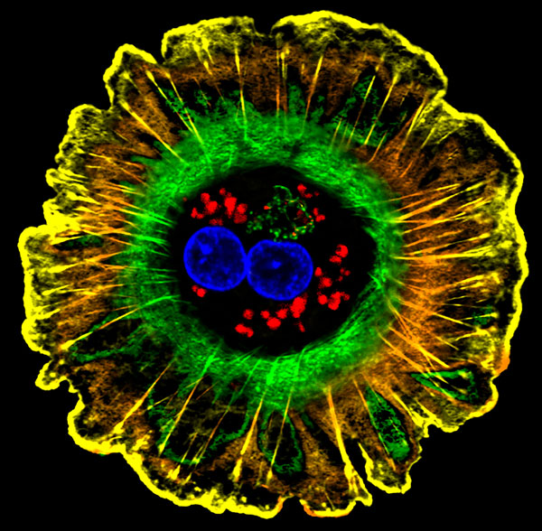
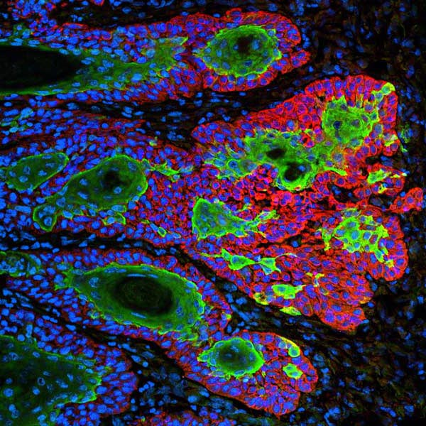 Skin Cancer Cells
Image credit: Markus Schober and Elaine Fuchs, The Rockefeller University, New York, N.Y.
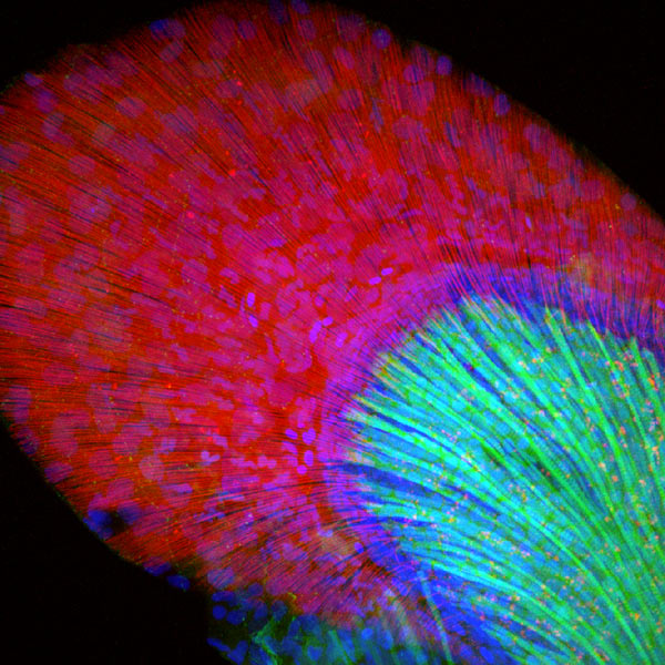 Developing Zebrafish Fin
Image credit: Jessica Plavicki, University of Wisconsin, Madison
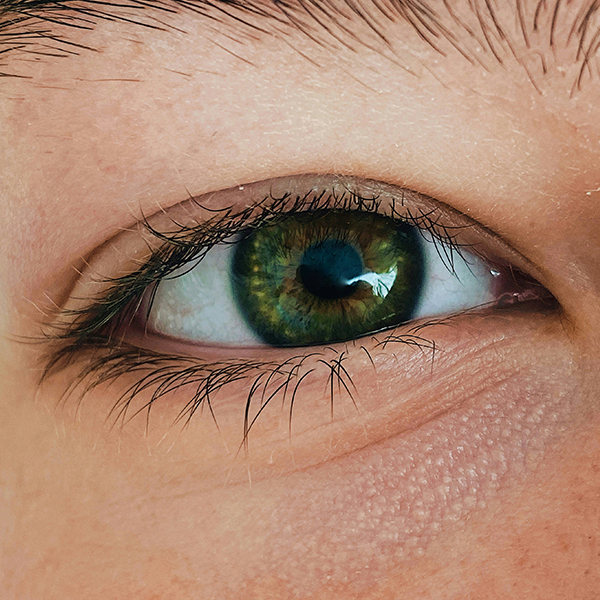
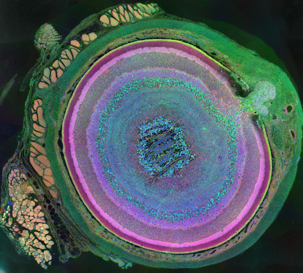 A Mammalian Eye Has Approximately 70 Different Cell Types
Image credit: Bryan William Jones and Robert E. Marc, University of Utah
Life: Magnified exhibition website
via [Phenomena: The Loom]